

(https://www.instagram.com/drjohn.orthovet)

Orthopedic Services
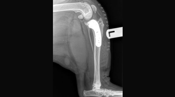
THR: Cementless Total Hip Replacement
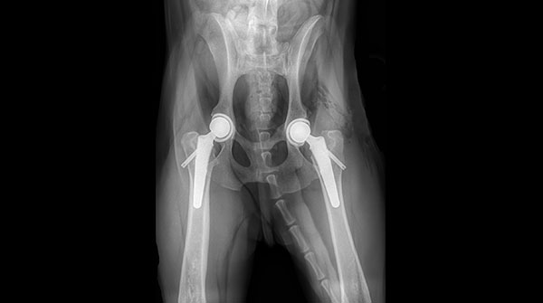
Dr. John performs Total Hip Replacements (THR) on dogs to relieve their pain, improve hip function and mobility and allow
them to return to an active, happy life. An implant replaces the arthritic joint, eliminating the
pain caused by bone rubbing against bone. This also restores the full range of motion of the limb.
Dogs with hip dysplasia over 9 months of age and weighing between 4 pounds - 170 pounds are likely candidates for THR.
Before committing your pet to surgery, we will perform a mandatory pre-operative exam to rule out other
problems. If your dog has undergone a femoral head excision, they will likely not be a good candidate for THR.
The overall goal of this surgery is to alleviate your pet’s pain, return them to normal hip function and improve
their quality of life. Other benefits include an increase in muscle mass, improved hip motion and increased
activity levels.
TPLO: Tibial Plateau Leveling Osteotomy
The procedure is based on the fact that the top part of the tibia bones normally sloped, resulting in tendency for the femur to
slide backward when the dog stands and puts weight on its knee. The Cranial Cruciate Ligament (CCL) normally holds the femur in place and prevents this motion. But when the CCL has ruptured, the femur can slide back and forth along the sloped tibia, when the dog is standing or walking. The continued motion contributes to pain and degeneration in the knee joint.
A solution to the knee joint instability would be to either replace the torn CCL, or remove the slope in the tibia. The TPLO does the latter: after making a cut in the top part of the tibia, the surgeon rotates this segment of bone until it is almost perpendicular to the ground. To allow the cut bone segments to heal, the tibia is then stabilized with a bone plate and screws. The result is that when your dog stand on its leg, the femur is resting on a flat tibia surface, and there is no longer the sliding motion in the knee.
For a video overview of the TPLO procedure, please see below video:
Arthroscopy
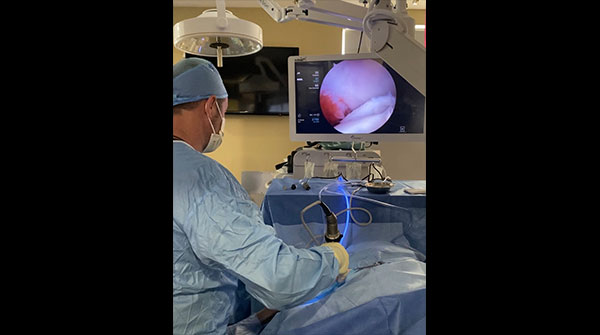
Arthroscopy is a minimally invasive surgical technique where a small camera is inserted into a joint in order to visualize and
diagnose joint problems. Once the joint has been evaluated, small instruments can also be
inserted into the joint in order to treat and repair certain types of joint damage. The benefits to an arthroscopic
procedure when compared to an open procedure include:
Arthroscopic procedures are generally performed on the elbow, shoulder, stifle (knee), and hock (ankle). We
utilize arthroscopy in the following procedures:
- Improved visualization of the joint to allow for better diagnostic accuracy
- Reduced scar tissue formation
- Faster recovery times
- Reduced rates of infection
- Decreased surgical pain due to smaller incisions and less tissue trauma
- Evaluating meniscal health
- Osteochondritis Dissecans (OCD)
- Fragmented Coronoid Process (FCP)
- Ununited Anconeal Process (UAP)
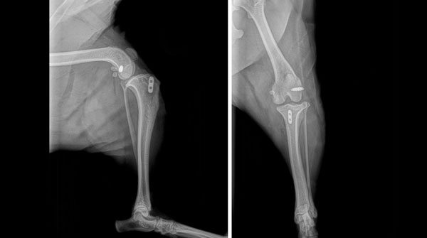
Total Elbow Replacement

Dr. John Brajkovich performs Total Medial Elbow Replacements (TMER) on dogs to relieve chronic elbow pain,
improve joint function, and help them return to a more comfortable, active life. An implant replaces the damaged joint surfaces, eliminating the pain caused by bone rubbing against bone and restoring smoother, more natural movement of the limb.
Dogs suffering from severe elbow arthritis or advanced elbow disease that has not responded to medical management may be candidates for TMER. Before committing your pet to surgery, we perform a pre-operative exam and advanced imaging to ensure there are no other underlying concerns and that this procedure is the right fit.
We use advanced 3D-printed, patient-specific guided systems to enhance surgical precision and consistency. These innovations help reduce surgical time, lower the risk of complications, and support a smoother recovery.
The overall goal of total elbow replacement is to alleviate your pet’s pain, restore comfortable joint function, and significantly improve their quality of life. Many patients experience improved limb use, increased strength, and a noticeable return to activity following recovery.
Extracapsular Cruciate Repair – Arthrex System
This technique was developed to provide a minimally invasive and improved method for extracapsular stabilization of the cranial
cruciate ligament (CCL). This technique does not require cutting of bone like the TPLO or TTA procedures. Instead, it uses small drill holes in the femur and tibia to pass a synthetic ligament-like biomaterial through a small incision to provide bone-to-bone stabilization during healing. The biomaterial used for the CCL repair is called FiberTape®️. This is a kevlar-like material that is used extensively in human surgery for many orthopaedic applications. This material has properties that make it stronger and less prone to failure than any other suture materials currently being used for CCL reconstructions.
TPLO / Patellar Luxation
This procedure combines a TPLO and a Patellar Luxation repair for patients who suffer from both a cranial cruciate ligament
tear and a patellar luxation in the same knee.
The procedure is based on the fact that the top part of the tibia bones normally slope, resulting in a tendency for the femur to slide backward when the dog stands and puts weight on its knee. The Cranial Cruciate Ligament (CCL) normally holds the femur in place and prevents this motion. But when the CCL has ruptured, the femur can slide back and forth along the sloped tibia, when the dog is standing or walking. The continued motion contributes to pain and degeneration in the knee joint.
A solution to the knee joint instability would be to either replace the torn CCL, or remove the slope in the tibia. The TPLO does the latter: after making a cut in the top part of the tibia, the surgeon rotates this segment of bone until it is almost perpendicular to the ground. To allow the cut bone segments to heal, the tibia is then stabilized with a bone plate and screws. The result is that when your dog stands on its leg, the femur is resting on a flat tibia surface, and there is no longer the sliding motion in the knee.
In addition to the TPLO procedure to remove the slope in the tibia to give stability back to the knee, spacers are placed to offset the TPLO plate allowing realignment of the patellar ligament. The patellar groove where the patella sits is also deepened to allow the patella to sit deeper in the groove.
Patellar Luxation
When the structures that make up your pet's knees (stifles) are misaligned or misshapen, a problem called a patellar luxation often
occurs. Your pet's kneecaps are an important component of a normally-functioning knee joint. These kneecaps (patella) are meant to ride in a groove on the face of the femur. The patella acts as a pulley, giving leverage to extend the knee as your pet walks. When a pet has a luxating (out of place) patella, this small bone jumps out of its normal grove as the leg is in motion. In over 90% of these cases in dogs, the patella jumps out of its tract to the inside of the pet's knee (medial patellar luxation or MPL). The signs you will see in your dog depend on how severe the problem is and how long the problem has been present.
Repair of patellar luxation involves three important steps
- Exploration of the knee joint to identify any ligament or cartilage damage.
- Realignment of the tibial tuberosity with the femoral groove.
- Formation of a normal femoral groove by deepening and widening it.
FCP: Fragmented Coronoid Process
The elbow joint is made up of three bones: the humerus (upper arm bone), radius and ulna (forearm bones). In this disease a small
portion of the ulna breaks off into the joint. This portion of the bone is called the medial coronoid process. FCP is the result of unequal growth rates of the radius and ulna, either individually or together. The joint space between the ulna and humerus is narrower than the joint space between the radius and humerus. The resulting uneven pressure on the medial coronoid process can develop cracks or fragments of the coronoid process.
Surgery or arthroscopy is generally indicated for your pet if he or she is lame or painful, and surgery and arthroscopy can stop or slow the progression of DJD. Surgery and arthroscopy tend to be more successful in younger patients (less than 12 months) and in patients that have not yet developed significant arthritis. Arthroscopy allows the surgeon to visualize the joint, remove the fragmented piece of bone and help smooth out any lesions in the joint. Surgery usually involves exposing the medial coronoid process via one of several methods and removal of the fragment.
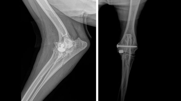
FHO: Femoral Head Osteotomy
A FHO is a surgical procedure that involves removal of the femoral head. The procedure is performed when hip joint disease
results in continuing pain and other methods of treatment are not feasible or did not produce the desired results. The procedure exposes the head section of the femur bone (the ball of the ball and socket joint), and then the head is removed using a small saw or a bone hammer and chisel. Rarely both sides are done in one operation, most times one side is done and allowed to heal before the other side is done. Unlike most other hip surgeries, the head of the femur is not replaced, When the femoral head is removed, the pain from pressure on the joint capsule and the cartilage is relieved. The patient forms what is called a pseudarthrosis, which is a fibrous (scar) tissue joint. The neck of the femur is usually removed at the same time as the head.
UAP: Ununited Anconeal Process
The anconeal process is a large piece of bone located at the growth plate at the top of the ulna. Normally, in
growing dogs, this area will close or fuse between 16-24 weeks of age. An ununited anconeal process is the failure of the anconeus to fuse with the ulna. Instability within the joint leads to damage of the articular cartilage, lameness and arthritis of the elbow joint. Surgical treatment is necessary to correct this condition and surgical excision is the most widely accepted method. In this procedure, the loose anconeal process is removed to prevent further irritation to the joint.
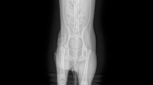
DPO: Double Pelvic Osteotomy
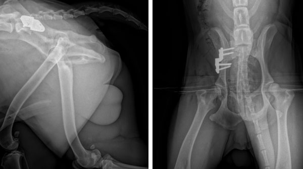
The goal of the DPO is to alter the dog’s natural hip joint and prevent the progression of arthritis. This procedure
involves cutting the pelvis in two places to allow the hip socket to be rotated at a predetermined
amount in order to fit a specially designed bone plate, thus improving the stability of the hip joint. Plate and
screws are used to keep the rotation in proper position.
Dogs experiencing complete luxation of the hip and grade IV hip dysplasia are not ideal candidates for the DPO procedure. The best candidates are younger dogs (between five and 10 months of age) with sufficiently deep hip sockets to accommodate rotation of the hip bone, and hip joints untouched by arthritis.
Dogs experiencing complete luxation of the hip and grade IV hip dysplasia are not ideal candidates for the DPO procedure. The best candidates are younger dogs (between five and 10 months of age) with sufficiently deep hip sockets to accommodate rotation of the hip bone, and hip joints untouched by arthritis.
PAUL: Proximal Abducting Ulnar Osteotomy
A PAUL surgery is performed on dogs that suffer from elbow dysplasia. This procedure involves making a controlled surgical
cut to the ulna and securing it in a new position with a plate and screw system. The surgery
is categorized as a load-altering osteotomy as it aims to unload the medial compartment of the elbow to
reduce pain and improve function and use of the limb.
Arthrodesis (Carpal and Tarsal)
Arthrodesis is a surgical fusing of a joint to eliminate movement in that part of a limb. An arthrodesis is generally performed
when there are no other options to save a joint and give a patient pain free function. To
perform an arthrodesis, the cartilage in the joint is removed and a bone graft is generally placed into the
spaces between the bones to promote fusion. The bones in the joint must then be rigidly stabilized using a
plate and screw system and sometimes an external fixation device. Following surgery a bandage or cast may
be applied to provide further immobilization of the joint. Activity is restricted for 8-12 weeks until there is
radiographic evidence that the bones in the joint have fused.
Angular Limb Deformities
Angular limb deformities can occur as a result of a genetic defect, in which the growth plates in long bones stop growing
prematurely in the animal’s development. This condition can cause severe problems with
mobility and function once the animal becomes unable to compensate for their deformity. Cats and dogs can
adapt to having a shortened limb, but their gait will be affected, and the adjacent joints will be put under
greater strain. Over time, this can lead to degenerative joint disease. We can correct these deformities by
sectioning and repositioning pieces of bone to correct the angular change.
Canine Unicompartmental Elbow (CUE)
Medial Compartment Disease (MCD) is the final stage of elbow dysplasia where the inside section of the joint collapses and
results in the grinding of bone on bone. To correct this, we can offer CUE surgery as a safe and
effective option for dogs that can no longer benefit from arthroscopic treatments and other non-surgical
alternatives.
With CUE surgery, we focus on the medial compartment. The CUE implant allows us to spare bone and provide a much less invasive alternative for resurfacing the medial compartment while also preserving the patient’s own cartilage in the lateral compartment. Overall, this procedure can reduce or even eliminate the pain and lameness associated with bone-on-bone contact.
With CUE surgery, we focus on the medial compartment. The CUE implant allows us to spare bone and provide a much less invasive alternative for resurfacing the medial compartment while also preserving the patient’s own cartilage in the lateral compartment. Overall, this procedure can reduce or even eliminate the pain and lameness associated with bone-on-bone contact.
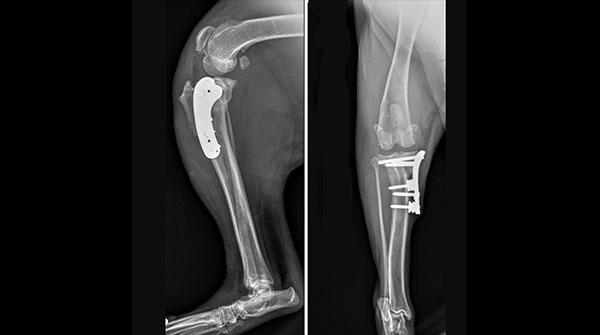
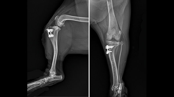
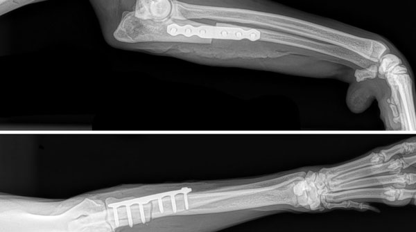
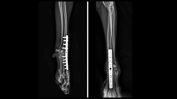
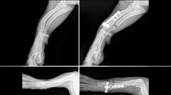
Fracture Repairs: Pinning, Plating & Internal or External Fixation

To repair a fracture, the ends of the bone must be brought together
and the continuity of the bone restored as close to normal as possible. This can be done with a closed technique—that is without exposing the bones—using traction and manipulation, trying not to disturb the natural healing processes already underway. Or, it can be done as an open technique, surgically exposing the bones by separating and, if necessary, cutting through muscle to visualize the fracture and to put it back together. The fracture must be immobilized to allow it to heal and this can be done in several ways.
External fixation describes the use of pins passed from outside the leg, through the skin and into the bones of the limb, ideally with at least three pins above and below the fracture. These pins can then connect to one another either by bars, or rods or cement or rings. External fixators can be applied open or closed, and combined with many other techniques making them extremely versatile.
Internal fixation describes the use of pins and wire, plate and screws. Plates and screws can be used for a variety of different fragments, but offer exceptionally stable fixation and in some cases the ability to squeeze or compress the ends of the bone fragments together. Such repairs can ensure an animal can be up and using a fractured limb as soon as possible.