Tracheal Stenting
Tracheal stenting is a surgical procedure designed to provide support to the cervical (neck) and thoracic (chest) portions of the trachea (Main airway of the body). This procedure is mostly used to
treat end stage conditions such as collapsing trachea. In collapsing trachea the rigid cartilage 'C' shaped rings of the trachea become soft, pliable and collapse with almost every breath- similar to if you have ever flattened a paper towel cardboard tube. This type of collapse makes it extremely difficult for the animal to take appropriate breaths in order to inflate their lungs. Tracheal stenting is the addition of a rigid-elastic material that provides structural support that mimics what normal healthy tracheal rings are designed to do. Advantages of stenting can include shortened anesthetic time compared to other tracheal surgical procedures, immediate improvement of clinical signs, and can be placed in the neck and chest regions in a noninvasive manner
Perineal Urethrostomy
This procedure can be done to both Cats and dogs, indications are animals that have chronic urinary obstructive problems or diseases that cause irreversible trauma and require reconstruction of the urogenital
system. This procedure is usually reserved for our male gendered cats and dogs. This procedure is most commonly done on male cats where as male dogs generally tend to have a scrotal urethrostomy rather than a perineal (space between the anus and scrotal sac). This procedure involves redirecting the outflow of urine through the urethra so that instead of the normal position through the penis where the urethra ends urination is now performed almost midway of the urethral length. So, why these locations? The perineum is where the urethra is at its widest point and most superficial (close to the outer skin layer) this makes it an ideal position to perform this surgery with the least amount of potential complications. Patients who undergo this surgery have almost immediate relief of their clinical problems and go back to normal life within 1-2 months post surgery. It is expected to see blood in the urine for this time but once the surgical site heals male cats who undergo this procedure manage quite well and return to normal life being able to urinate with no problems.
Perineal Hernia
Perineal hernia occurs due to weakness and separation of the muscle groups that make up the 'pelvic diaphragm'. These muscles are located on either side of the tail down to the ventral (bottom) most border
of the anus. This weakness can cause abdominal organs to protrude or become trapped with the weakened spaced. Organs like the rectum, prostate, bladder and intestines. This weakness usually occurs in older animals 7-13- most occurring from 7-9 of both small and large dogs breeds and almost exclusively in male intact dogs, comprising about 83-93% of all cases. We are still not 100% what causes this herniation to occur but we seem to think that short docked tail animals, genetic predisposition, and male androgens ie. Testosterone all have an effect; and when dogs were castrated it reduced recurrence form 43% to 23%. The hernia is repaired by creating a new pelvic diaphragm by re-orientating certain muscles in the area to reinforce the current weakened muscles present. There are many different types of reinforcement techniques and each one has its pros and cons. The technique that is done is based on individual considerations and how severe the hernia is. This surgery is tailored to your animals needs to ensure the best outcome with minimal complications.
Brachycephalic Airway Syndrome

Brachycephalic Airway Syndrome is a combination of anatomical formation of the dogs airway structures that interferes with normal breathing and taking in air to the lungs. This syndrome affected only
brachycephalics (short/squished/scrunched face dogs) ie. Bulldogs, pugs, french bulldogs. The 3 main components are stenotic nares (more closed nostrils), elongated soft palate and a hypoplastic trachea (causing narrowing of the main airway). Another abnormalities can be everted laryngeal saccules which are 'pouches' that live on either side of the opening of the trachea and in severe cases the 'pouches' evert and block portions of the trachea further hindering airflow into the lungs. The two main procedures to combat this syndrome are widening the nostril by cutting a portion of the tissue away in the hopes to widen the airflow. Every increase in nostril opening increased airflow x4. So even a small enlargement goes a very long way. A soft palate resection is also done to prevent epilotal (the flap covering the trachea when swelling) entrapment so that breathing can be done with both nose and mouth unhindered. Because these dogs can sometimes have other pathologist that go along with this syndrome, a comprehensive physical exam must be done prior to surgery, bloodwork, starting on reflux/vomiting medications and +/- radiographs should be done to ensure minimal complications during surgery and postoperatively.
Cherry Eye Repair
You may have heard the term 'Cherry eye' before or have had animals who suffer from this condition. Cherry eye is a condition in which the nictitating membrane, more commonly known as the third
eyelid, protrudes out of its normal location-underneath the lower lid in the inner corner of the eye. This third eyelid houses a gland that is responsible for producing up to 35% of the tear film in dogs. Due to this large production, cherry eye repair is vital in maintaining a healthy environment for the eye. Cherry eye repair is most commonly done surgically, for very mild cases massaging can be done daily to try and 'pop' the gland back into place but that technique seldom works. Surgical techniques are designed to create a new pocket for the gland to sit in so it can stay protected and continue to function normally.
TECA: Total Ear Canal Ablation & Bulla Osteotomy
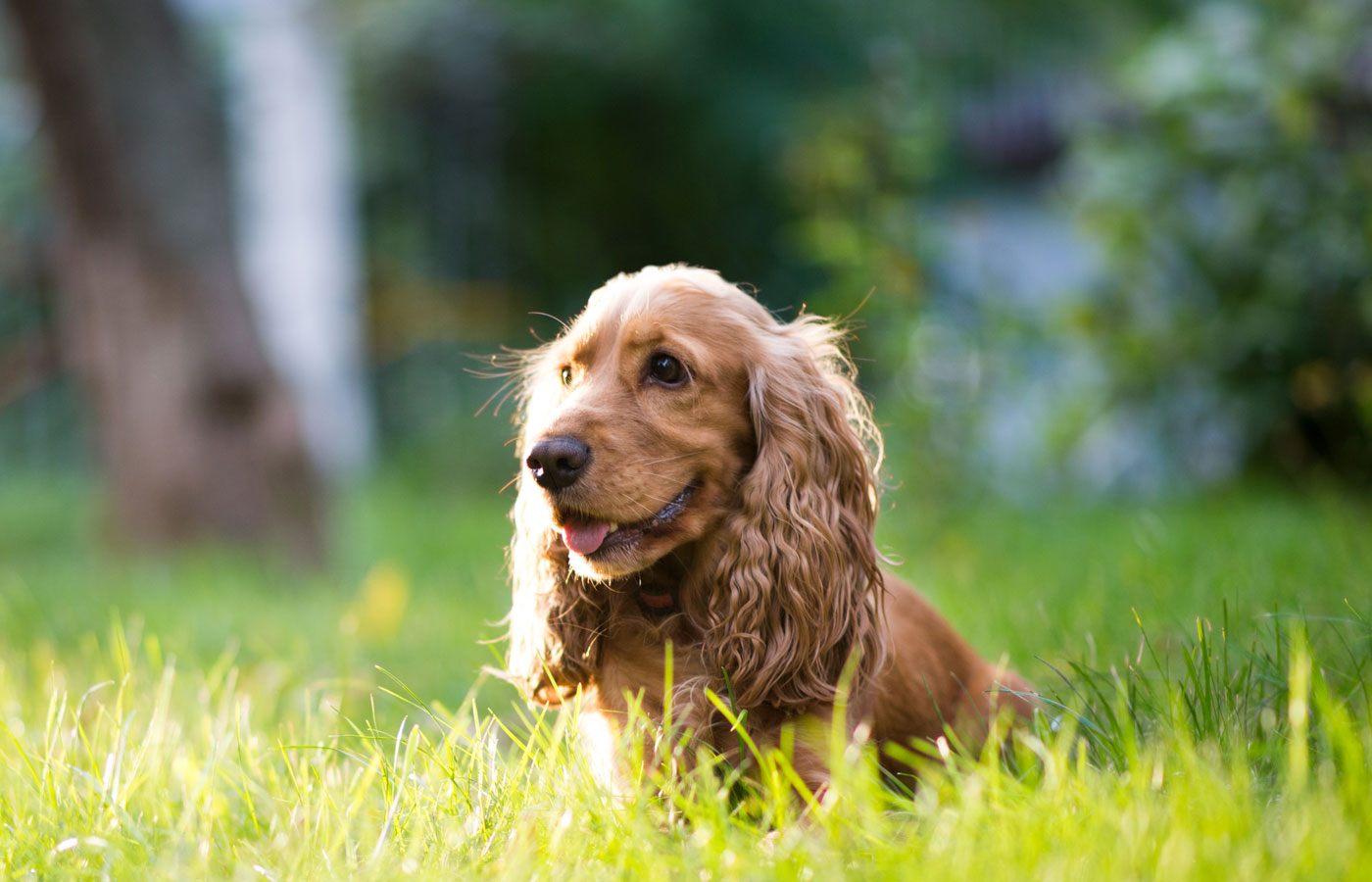
Total Ear Canal Ablation & Bulla Osteotomy or TECA-BO for short is usually a procedure done for end stage inflammatory diseases such as infections, neoplasia (cancer), trauma, stenosis (narrowing)
or obstructive issues (ie. polyps of the ear canal). The procedure involves removal of the outer ear canal and tympanic bulla (a bone shaped cavity that can accumulate fluid and debris and abnormal tissue). Think of the ear canal as 'L' shaped with a ball the end (tympanic bulla). The surgical procedure is removing the 'L' portion (the canal) and making a hole in the ball at the end to facilitate draining and removal of abnormal tissue. The most common complication with this surgery is hearing loss, nerve damage ie. Horner's syndrome (more so in cats than dogs), and facial nerve damage, this is because the nerves that innervate the face cross the surface of the bony prominence of the ear. Great care is always taken during this procedure to minimize trauma to these structures and nerves but if the disease process is severe enough these complications may arise.
Hernia Repairs

A hernia is classified as an abnormal exit of any tissue or organ. The most common type of hernia seen is an umbilical hernia where most of the time abdominal fat is herniated through the umbilical
cord attachment due to not closing properly after birth. The biggest risk of herniation is organ herniation, where a segment or the entire organ exits its original anatomical location into the herniated space. As a general rule of thumb every hernia should be treated due to the potential risk of organ herniation. Traumatic hernias are always repaired because there is always an organ herniated. The most severe herniation is a diaphragmatic hernia where abdominal contents pass through the abdomen into the chest. These are life threatening and are always repaired. These types of hernias are generally repaired at a specialist center due to requiring overnight care and positive pressure ventilation due to compression of the lungs. If your dog or cat does have a hernia, general recommendations are to come in for a comprehensive physical exam for assessment and going
forward from there.
Complex Tumor Resections
Tumors come in a variety of shapes, sizes and can affect every kind of tissue in the body. Some tumors removals can be straightforward and uncomplicated such as small to medium and
some large skin masses, eyelid masses, and lipomas etc. But some can be very tricky require advanced reconstructive surgeries such as skin flaps, mesh wiring and removal of organs. Some complex tumor sadly cannot be removed and attempting so can be life threatening to animals. This is why every mass found on your animal should be seen for a comprehensive physical exam, the masses tested to see what kind of tissue is evolved and imaging such as radiographs (x-rays), CT scan or even in some cases MRI. Based on cell type, size and anatomical position of the mass a surgical plan will be devised and surgery schedules. Upon removal of any mass, it is highly recommended the mass sent off for histopathology to determine the exact type of tumor, grade and to assess for tumor free margins.
Anal Gland Excision

This procedure involves the complete removal of the anal gland or sac when they do not function properly due to various conditions/complication.
The most common diseases we see affecting the anal gland are:
While some conditions do not require the anal gland to be removed, others most certainly do. Anal sac cancers are diseases that will always lead to the removal of the anal gland. Apocrine gland adenocarcinoma (anal gland/sac cancer) makes up nearly 20% of all perianal cancers and about 2% of all skin and subcutaneous (under the skin) cancers/masses. Surgical excision of these glands do not affect the quality of life of the animal and they do not serve a vital function for day to day life. Baring any complication, anal gland excision is the best way to remove a chronic or potential life threatening conditions.
The most common diseases we see affecting the anal gland are:
- Impaction
- Sacculitis (inflammation of the anal gland)
- Abscessation
- Neoplasia (cancer)

